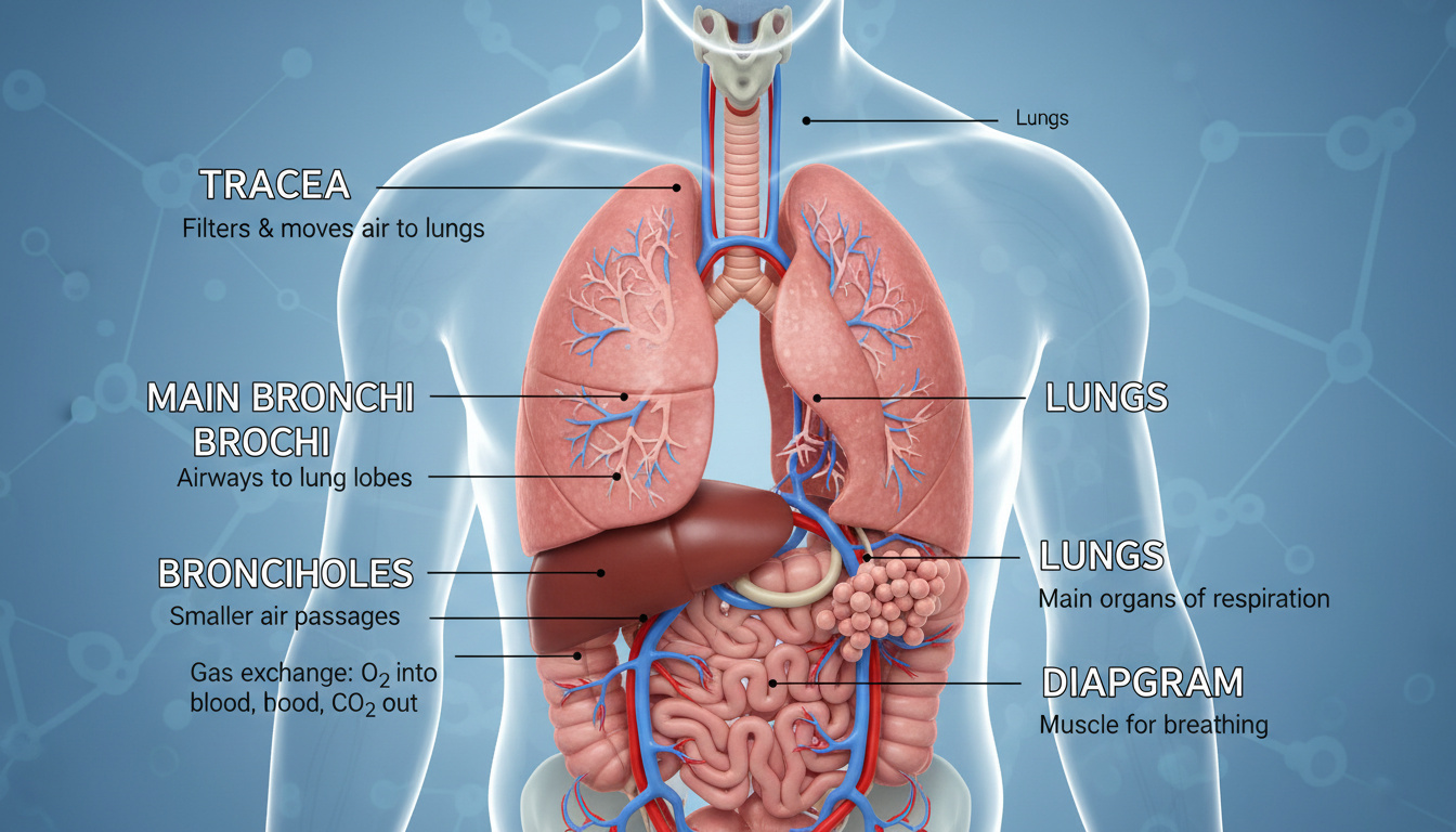
Accurate anatomical diagrams serve as foundational tools for understanding human physiology. The respiratory system, comprising intricate pathways for air conduction and gas exchange, requires precise visual representation to clarify the relationships between the nasal cavity, bronchial tree, and pulmonary structures. Students, educators, and medical professionals rely on labeled schematics to distinguish between the upper and lower respiratory tracts, with authoritative resources such as the National Heart, Lung, and Blood Institute providing standard anatomical references.
While nutritional support such as Bone Broth Benefits may contribute to overall wellness, anatomical literacy remains essential for comprehending pulmonary function. Standard diagrams typically illustrate airflow from the nostrils through the nasopharynx, larynx, trachea, and bronchi to the alveoli. These visual aids demystify the spatial organization of organs that facilitate oxygen transport and carbon dioxide elimination.
Contemporary medical education utilizes both static illustrations and three-dimensional models to convey structural complexity. High-fidelity representations help learners identify the C-shaped cartilage rings of the trachea, the branching patterns of bronchioles, and the segmental divisions within the right and left lungs.
What Are the Main Parts of the Respiratory System?
| Organ | Location | Function | Diagram Reference |
|---|---|---|---|
| Nose/Nasal Cavity | Upper tract, superior | Filters, warms, humidifies air | Sagittal view anterior |
| Trachea | Lower tract start | Air conduit with cartilage rings | Coronal section |
| Bronchi | Within lungs | Primary to tertiary branching | Bronchial tree schema |
| Alveoli | Lower tract core | Gas exchange via capillary networks | Histological cross-section |
- The conducting zone extends from the nose to terminal bronchioles without participating in gas exchange.
- The respiratory zone begins at respiratory bronchioles, encompassing microscopic alveolar sacs.
- Right lung contains three lobes with ten bronchopulmonary segments; left lung contains two lobes with eight segments.
- Air filtration occurs primarily in the nasal cavity’s mucosal lining and paranasal sinuses.
- The larynx contains nine cartilages and serves dual functions in airway protection and phonation.
- Bronchioles less than one millimeter in diameter mark the transition from conductive to respiratory zones.
- The pleura forms a protective serous membrane enclosing each lung within the thoracic cavity.
| Structure | Primary Role | Key Fact |
|---|---|---|
| Nose/Nasal Cavity | Air filtration, warming | Lined with ciliated pseudostratified mucosa |
| Paranasal Sinuses | Resonance, humidification | Connected to nasal cavity via ostia |
| Pharynx | Passageway | Three divisions: naso-, oro-, laryngopharynx |
| Larynx | Airway protection, voice | Nine cartilages including thyroid and cricoid |
| Trachea | Windpipe | Ten to twelve centimeter tube with C-shaped cartilage rings |
| Primary Bronchi | Main branch | Right bronchus wider, shorter, more vertical than left |
| Secondary Bronchi | Lobar supply | Three on right, two on left corresponding to lobes |
| Bronchioles | Distribution | Lack cartilage, smooth muscle regulated |
| Terminal Bronchioles | End of conducting zone | No gas exchange occurs here |
| Respiratory Bronchioles | Start of respiratory zone | Sparse alveoli appear on walls |
| Alveolar Ducts | Gas exchange pathway | Lined with alveoli and alveolar sacs |
| Alveoli | Gas exchange | Surrounded by capillary networks; site of O₂ and CO₂ transfer |
How Does the Respiratory System Work?
The Mechanics of Pulmonary Ventilation
Inhalation and exhalation depend upon pressure changes within the thoracic cavity. The diaphragm contracts during inspiration, increasing thoracic volume and drawing air through the conducting passages. Exhalation typically proceeds passively as the diaphragm relaxes, though forced expiration engages accessory muscles. The MedlinePlus respiratory system overview describes this process as the mechanical foundation for external respiration.
Gas Exchange at the Alveolar Level
Oxygen diffusion occurs across the respiratory membrane where alveoli interface with pulmonary capillaries. Hemoglobin in red blood cells binds oxygen while carbon dioxide exits the bloodstream to be expelled. This exchange requires the extensive surface area provided by alveolar clustering within pulmonary lobules.
Air travels unidirectionally through the respiratory tract. The conducting zone transports, cleanses, warms, and humidifies air without gas exchange, while the respiratory zone handles actual oxygen and carbon dioxide transfer across capillary networks.
What Are the Upper and Lower Respiratory Tracts?
Upper Respiratory Structures
The upper tract encompasses the nose, nasal cavity with paranasal sinuses, and the pharynx divided into nasopharynx, oropharynx, and laryngopharynx. These structures moisten, warm, and filter incoming air while routing it toward the lower passages. The larynx, containing nine distinct cartilages, marks the transition point where the respiratory and digestive pathways diverge, protected by the epiglottis during swallowing.
Lower Respiratory Pathways
Beginning at the larynx, the lower tract includes the trachea with its C-shaped cartilage rings, the primary bronchi, and the extensive bronchial tree. The right main bronchus remains wider and more vertical than its left counterpart, explaining why aspirated foreign objects more commonly enter the right lung. Subsequent divisions into lobar and segmental bronchi eventually terminate in bronchioles and alveolar structures where gas exchange occurs. Mayo Clinic multimedia resources illustrate these branching patterns in clinical contexts.
The pleura forms a protective serous membrane enclosing each lung within the thoracic cavity. This double-layered structure reduces friction during respiratory movements and maintains negative pressure essential for lung expansion.
How Does Air Flow Through the Respiratory System?
- Inhalation: Diaphragm contracts, thoracic volume increases, air enters via nostrils.
- Nasal Conditioning: Air passes through nasal cavity, warming and humidifying to body temperature and saturation.
- Pharyngeal Passage: Air moves through nasopharynx and oropharynx into the laryngopharynx.
- Laryngeal Routing: Epiglottis prevents food entry; vocal cords adjust; air enters trachea.
- Tracheal Descent: Air travels through the ten to twelve centimeter trachea supported by cartilage rings.
- Bronchial Branching: Primary bronchi divide into lobar (secondary) then segmental (tertiary) bronchi.
- Bronchiolar Transition: Airways narrow to less than one millimeter diameter, becoming terminal bronchioles.
- Respiratory Zone Entry: Air reaches respiratory bronchioles where alveoli begin appearing on walls.
- Alveolar Exchange: Oxygen diffuses across alveolar-capillary membrane into blood; carbon dioxide exits.
- Exhalation: Diaphragm relaxes, thoracic volume decreases, carbon dioxide-rich air expelled.
What Is Established Versus Variable in Respiratory Anatomy?
| Established Anatomical Facts | Areas of Individual Variation |
|---|---|
| The division between upper and lower tracts occurs at the larynx. | Exact number of segmental bronchi varies between eight and ten in different anatomical classifications. |
| Right lung has three lobes; left has two. | Tracheal length varies between ten and twelve centimeters among individuals. |
| The conducting zone contains no alveoli; gas exchange occurs only in the respiratory zone. | Accessory or ectopic bronchi occur in rare anatomical variants not detailed in standard diagrams. |
| Alveoli serve as the exclusive sites for oxygen and carbon dioxide exchange with blood. | Total alveolar surface area varies significantly based on body size and pulmonary health status. |
Why Accurate Diagrams Matter in Medical Education
Visual literacy in respiratory anatomy underpins clinical diagnostics and surgical planning. Misidentification of bronchopulmonary segments can lead to errors in interpreting radiological scans or planning resections. Anatomical diagrams provide the spatial framework necessary for understanding pathologies such as pneumonia distribution or tumor localization.
Research from physiological studies confirms that comprehension of the conducting and respiratory zones requires understanding their distinct epithelial linings and functional boundaries. Furthermore, evidence-based nutritional support, detailed in Bone Broth Benefits – Evidence-Based Facts, highlights the importance of maintaining respiratory tissue integrity through dietary components.
Medical References and Anatomical Authority
The bronchial tree schema demonstrates hierarchical segmentation from trachea through primary bronchi to lobar, segmental, and subsegmental divisions, terminating in alveolar structures where gas exchange occurs.
— 3D Modeling of Respiratory Organs, Web3D Consortium
The upper respiratory tract functions primarily in air conduction, warming, humidification, and filtration, while the lower tract serves as the apparatus for pulmonary ventilation and external respiration.
— Respiratory Anatomy Lecture Notes, San Diego Miramar College
Interactive atlas views allow rotation to visualize the tracheal bifurcation, right and left main bronchi, and the distinct lobar organization of the pulmonary parenchyma.
— Atlas Lab Manual, Visible Body
Essential Points About Respiratory System Diagrams
Comprehensive respiratory diagrams illustrate the anatomical continuum from nasal cavity to alveoli, distinguishing between the air-conducting upper tract and gas-exchanging lower tract. Accurate labeling of the bronchial tree, segmental divisions, and pleural boundaries remains essential for medical education and clinical practice, supported by authoritative anatomical references and three-dimensional modeling resources.
Frequently Asked Questions
Where can I find a respiratory system diagram for class 10?
Educational resources from state education departments provide labeled diagrams suitable for secondary school curricula, showing nostrils through alveoli with simplified bronchial branching.
Are 3D respiratory system diagrams available online?
The Web3D Consortium publishes hierarchical joint models and animation schemas, though fully interactive web-based 3D viewers require specialized anatomical atlases.
What is the primary function of the respiratory system?
Gas exchange constitutes the core function: oxygen enters blood while carbon dioxide exits via alveolar-capillary networks, facilitated by the conducting zone which prepares air without direct diffusion.
How many lobes does each lung have?
The right lung contains three lobes with ten bronchopulmonary segments, while the left lung has two lobes containing eight segments.
What separates the upper and lower respiratory tracts?
The larynx marks the anatomical boundary; structures above it comprise the upper tract, while the trachea and below form the lower tract where gas exchange occurs.
Can I download a printable PDF of the respiratory system?
Institutional repositories such as Pearson higher education offer downloadable diagrams showing upper and lower tract divisions with labeled structures.
What are alveoli and why are they important?
Alveoli are microscopic air sacs surrounded by capillary networks where oxygen diffusion into blood and carbon dioxide removal occur through thin respiratory membranes.
Does the trachea contain bone?
The trachea contains C-shaped cartilage rings composed of hyaline cartilage, providing rigid support to prevent airway collapse while allowing flexibility for neck movement.
What educational resources explain respiratory anatomy interactively?
Khan Academy provides video tutorials and interactive lessons covering respiratory system biology for various educational levels.
Rachel Walsh leads fact-checking, source verification and corrections at Aussie Pulse.